View Glossary
Ocular disease is common and may be due to injuries, systemic health, breed, or genetics. Some of the most common eye conditions that we diagnose and treat are listed below.
Eyelid abnormalities
- Entropion and ectropion
- Ectopic cilia
- Distichiasis
- Eyelid tumors
- Trichiasis
- Eyelid agenesis
Prolapsed gland of the third eyelid (cherry eye)
Keratoconjunctivitis (Dry Eye)
Corneal disease
- Corneal ulcer
- Indolent corneal ulcer
- Descemetocele
- Corneal sequestrum
- Feline herpesvirus keratitis
- Chronic superficial keratitis (pannus)
Anterior uveitis
Glaucoma
Lens Disease
- Cataract
- Lens luxation
Orbital tumors and abscesses
Intraocular tumors
Retinal disease
- Retinal detachment
- Retinal degeneration
- Retinal dysplasia
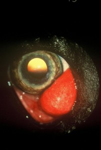
Cherry Eye
When the tear gland of the third eyelid pops out of position, it protrudes from behind the eyelid as a reddish mass. This prolapsed tear gland condition is commonly referred to as “cherry eye”. The problem is seen primarily in young dogs, and often in certain breeds such as the Cocker Spaniel, Lhasa Apso, Shih-Tzu, Poodle, and Bulldog.
Despite its appearance, cherry eye itself is not a painful condition. However, the longer the tear gland is exposed, the more likely it will come irritated and inflamed. This tear gland makes 30-50% of the tear film. When the gland is prolapsed, the small tear ducts do not function normally (think of a “kinked” garden hose).
To correct cherry eye, surgical REPLACEMENT of the gland is necessary. This treatment is superior to a somewhat older technique of surgically REMOVING the gland. The gland of the third eyelid plays an important role in maintaining normal tear production. We now know that dogs that have had the tear gland removed are predisposed to developing Dry Eye Syndrome later in life. Dry Eye Syndrome is uncomfortable for the patient, and requires the owner to administer topical medications several times a day for the remainder of the patient’s life.
Entropion
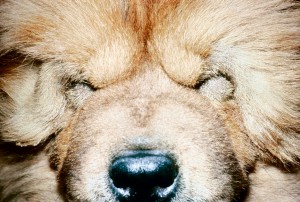
Entropion is an inward rolling of the eyelid. The problem is most common in the Shar Pei, Chow Chow, Bulldog, Retrievers, Rottweiler, and Setters. Although the exact genetic pattern is usually not known, the problem is most likely caused by many genes that are responsible for the overall head and face conformation. Therefore, we recommend that you do not breed your pet since the problem can be passed to the puppies.
When the eyelids roll in, the hair on the outside of the lid rubs on the surface of the eye and cause corneal irritation. In some patients, entropion leads to corneal ulceration. Common symptoms associated with entropion include squinting, tearing, and rubbing.
The treatment for entropion is surgery to remove excess skin and muscle along the eyelid margin. This is cosmetic surgery and after reconstructing the eyelid, the lids should look normal.
Eyelid Tumor
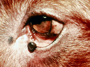
Eyelid tumors come in many sizes and shapes. Some are small and slow-growing, while others may rapidly grow in size. Some lid tumors are not bothersome, while others may rub against and irritate the surface of the eye, or even obstruct vision.
Small, slow-growing tumors that do not cause other ocular problems and are not of a type suspected to be malignant often do not need to be removed. On the other hand, large or fast-growing tumors that cause irritation, obstruct vision or periodically bleed are often surgically removed.
Cataract
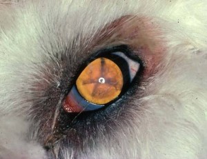
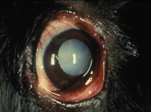
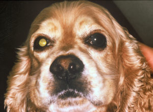
Cataracts are opacities in the lens of the eye. Many people mistakenly think the cloudiness is on the surface (thought to be a “film” on the eye), but in fact, the cloudy lens is deep inside your pet’s eyeball.
Why Did My Pet Get Cataracts?
Most cataracts are inherited, and are found in many breeds such as the Cocker Spaniel, Poodle, Husky, Schnauzer, Bichon Frise, Golden and Labrador Retrievers, and terriers. Other causes of cataracts include: Diabetes, trauma, inflammation, and puppy milk replacers. Many cataracts will worsen to the point of blindness but certain types, especially in the Retriever breeds, can remain small for the entire life of the patient. A common phenomenon occurs in many developing cataracts where the patient can develop an allergic type of reaction to the cataract. This allergic reaction is a LOCAL reaction and can result in many complications such as scar formation and glaucoma.
How Are Cataracts Treated?
Treatment for cataracts is surgical removal by a technique called phacoemulsification. Surgery may be performed in one or both eyes depending on the specifics of each patient. Before surgery is performed, your pet will have two special tests (electrogretinogram and ocular ultrasound) beyond the full eye exam to check the health of the retina or nerve layer in the back of the eye. The Animal Eye Center of New Jersey was the first veterinary hospital in the world to utilize the Whitestar Signature Phacoemulsification system. The Whitestar Signature Phacoemulsification System is one the most advanced cataract removal systems available for people and we employ this technology at the Animal Eye Center of New Jersey to restore your pet’s vision! Foldable intraocular lens implants are placed inside the eyes after cataract surgery to help focus vision.
Corneal Ulcer
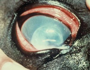
Similar to a cut or scrape on your skin, an ulcer is an unprotected wound on the surface of the eye. The normal cornea is covered by a layer of tissue called the epithelium, sort of like “skin” over the deeper eye layers. When the epithelium is damaged, infections can occur and result in complete perforation of the eye if left untreated. Clinical signs of corneal ulcer include squinting, redness, cloudiness, tearing, and lethargy. A special green stain can be used to highlight the ulcer on the cornea. There are many causes of corneal ulcer such as injuries, abnormal eyelashes that irritate the surface, lack of tear production, infections, and many types that we don’t understand very well.
Corneal ulcers are characterized according to location, depth, associated diseases, and cause. The treatment of the ulcer depends on the type and depth of the ulcer. Some corneal ulcers require simple application of medications to prevent infection and alleviate pain, whereas very deep corneal ulcers require surgically placed grafts to prevent complete perforation of the eye.
Corneal Dystrophy
The term Corneal Dystrophy is used to describe a variety of disorders affecting the layers which make up the cornea (the clear surface of the eye). Cloudiness is the primary characteristic of corneal dystrophy that you are likely to notice. In some cases, the dystrophic (cloudy) areas of the cornea are prone to intermittent erosions which could cause your pet to squint from time to time. Specific topical eye ointments can be used to create a protective layer over dystrophic areas, reducing the chance that corneal erosions will occur.
Corneal dystrophy tends to run in certain lines or breeds of dogs, including the Boston Terrier, Sheltie, Samoyed, Husky, and Corgi. Once corneal dystrophy occurs, the cloudy dystrophic areas will not go away. Vision is usually not affected by small areas of corneal dystrophy, but larger cloudy areas can sometimes interfere with normal vision.
Glaucoma
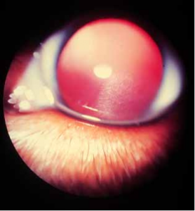
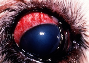
Glaucoma is defined as “an increase in pressure in the eye, with a loss of vision.” The disease is quite painful in most cases, especially when the eye pressure is very elevated. The signs of glaucoma include redness, cloudy eye, tearing, squinting, loss of vision, an enlarged eyeball, unusual aggressiveness, lethargy, and loss of appetite. The normal physiology of the fluid in the eye calls for the fluid to be made in one structure behind the pupil (ciliary body), travel through the pupil, and exit out the space between the cornea and the iris. When the fluid cannot properly drain from the eye, the pressure in the eye is increased. An analogy would be a kitchen sink — if the drain is open and the water is running, there is no problem. However, if a plug is placed in the drain and the water keeps coming, then the sink fills up with water and overflows! Some patients have primary glaucoma where there is no concurrent disease but some secondary causes of glaucoma include inflammation, trauma, and tumors. All of these factors can obstruct the drainage of fluid from the eye. Unfortunately, there is no “cure” for glaucoma. It is a disease that is managed with medical therapy or surgery.
Glaucoma is an ophthalmic emergency and must be treated immediately. If the pressure remains elevated for even a few hours, permanent vision loss occurs. The disease is difficult to treat but several option are available depending on whether the patient still has vision, the species of the patient, financial considerations, etc. Some of the options include medical management with pills and eye drops, shunt and laser treatment to reduce the fluid production and improve outflow, and removal of the blind and painful eye with a cosmetic prosthesis or complete removal.
Intraocular Prosthesis
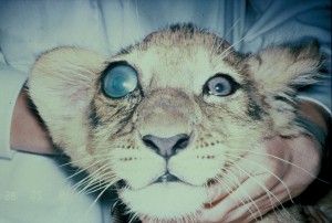
Cosmetic Prosthesis Implant- This surgical option is used only on a blind eye. Under general anesthesia, the inner contents of the eye are removed and replaced with a black silicone ball. The patient keeps the outside of the eye, including the eyelids, cornea, sclera and extraocular muscles, so that the eye moves and looks fairly normal. Instead of the colored iris, the eye usually looks silvery grey or black, but can be white depending on the amount of cloudiness of the cornea before surgery. The advantage of the procedure is that once the contents of the eye are removed, there is no more painful glaucoma to treat or worry about in that eye. Furthermore, the removal leaves the patient looking cosmetically more “normal” than enucleation.
Uveitis
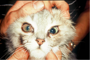
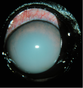
Uveitis refers to an inflammation (irritation) of the part of the eye that supplies blood to the eye. If the eye is to remain clear so that light rays can focus on the back of the eye, the blood vessels must be located out of the way of the clear structures such as the cornea and lens. When the blood vessels become irritated, blood cells and debris leak out and result in cloudiness. Clinical signs of uveitis include: cloudiness, redness, tearing, squinting, bleeding into the eye, and loss of vision.
There are many causes of uveitis such as trauma or cataract formation. Many types of infections and tumors can cause uveitis in the dog and cat. Some of the infections in dogs include: Rocky Mountain spotted fever, Ehrlichiosis, infected uterus in females, hepatitis virus, and systemic fungal infections and possibly Lyme’s disease and Anaplasmosis. In cats the causes can include: feline leukemia virus, feline AIDS virus, FIP virus, Bartonellosis and Toxoplasmosis. However, many patients with uveitis DO NOT have an obvious underlying cause. We evaluate the entire patient to decide whether specific laboratory testing is necessary to search for a cause of the uveitis.
Uveitis can result in several eye complications such as cataract formation, scar tissue, retina disease, and glaucoma. Treatment for uveitis is aimed at reducing the inflammation and preventing the complications. The treatment protocol will vary for each patient but often includes a steroid eye medicine to decrease the inflammation, atropine to alleviate the pain (people with uveitis get a tremendous headache!), oral anti-inflammatory medications to decrease inflammation, and possibly medicine to prevent glaucoma.
Retinal Detachment
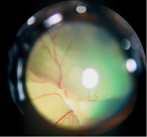
The retina is the nervous layer of the eye, and it normally lies flat against the back of the eye. The retina is responsible for collecting light impulses which are then transferred to the brain and interpreted as vision. When the retinas detach from their normal position, they cease to function and the patient becomes blind.
Retinal detachment can be caused by tumors, fungal infections, severe trauma, inflammation, genetic predisposition, high blood pressure, or immunity problems. Depending on what caused the detachment and how long the retina has been detached, in some cases the retinas can be reattached and vision restored. In many cases, however, the retina deteriorates and the prognosis for vision is poor. Early intervention (diagnosis and treatment) is critical with retinal detachment in order to help save vision.
Hyphema
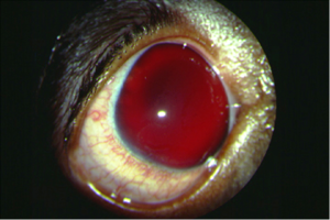
Hyphema refers to bleeding inside the eye. The condition can be caused by a variety of factors, including trauma, tumor, clotting disorders, multiple eye diseases, or high blood pressure. Hyphema usually causes secondary inflammation inside the eye (uveitis). Sometimes as the blood clots and settles it can plug the drainage ducts inside the eye and cause glaucoma. Treatment for Hyphema depends on the overall health of the eye. Often topical steroids are used to decrease secondary inflammation.
Retinal Degeneration
The retina is the nervous layer in the back of the eye responsible for collecting and transmitting light images to the brain where they can be interpreted as vision. Retinal Atrophy and Retinal Degeneration are general terms used to describe areas of the retina that are functioning improperly or are “worn out”. Small areas of degeneration may not cause vision problems, but large areas of degeneration can significantly interfere with normal vision. Retinal degeneration or atrophy can result from severe eye trauma, infection, or retinal detachment. Treating the underlying cause will prevent further degeneration and vision loss. Unfortunately, there is no way to repair the areas that are already damaged.
